Cell Adhesion Studies Using the Rheoscope
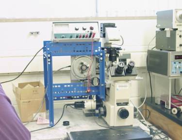
The Rheoscope is a cone plate viscometer used in conjunction with an inverted microscope. It is used to shear cells in a controlled manner that are either attached to the bottom plate or suspended in a medium. The advantages of this system are that the shear rate can be precisely controlled and that the cellular response to that shear can be seen through the microscope or on video. The rheoscope is shown at right.
Colon Cancer cell adhesion assays are currently being
conducted using the rheoscope. Focal
Adhesion Kinase is a kinase that has proven to play an important role in
colon cancer cell differentiation and metastasis.
These studies are being used to determine if Focal Adhesion Kinase
affects cell detachment.
Figure 1. FAK containing cells
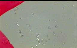
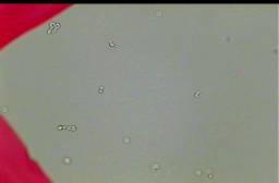
Figure 2. Cells not containing FAK
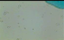
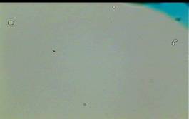
Figure 3. Video of a cell detaching from shear experienced in the rheoscope.
(Click on image below to view video)
Delaney, Melissa. The effect of focal adhesion kinase (FAK) on FEK 293 cell line (FEK 293) cell detachment. Thesis. 2003.